Endodontics

Why does visibility matter in microscope-assisted endodontics?

Dr. Onur Demir, DDS, PhD · 4 min read · Updated 12 September 2026

Root canal systems can contain narrow entrances, additional anatomy, calcification and changes caused by previous treatment. Magnification and coaxial illumination can make these details easier to identify and manage.

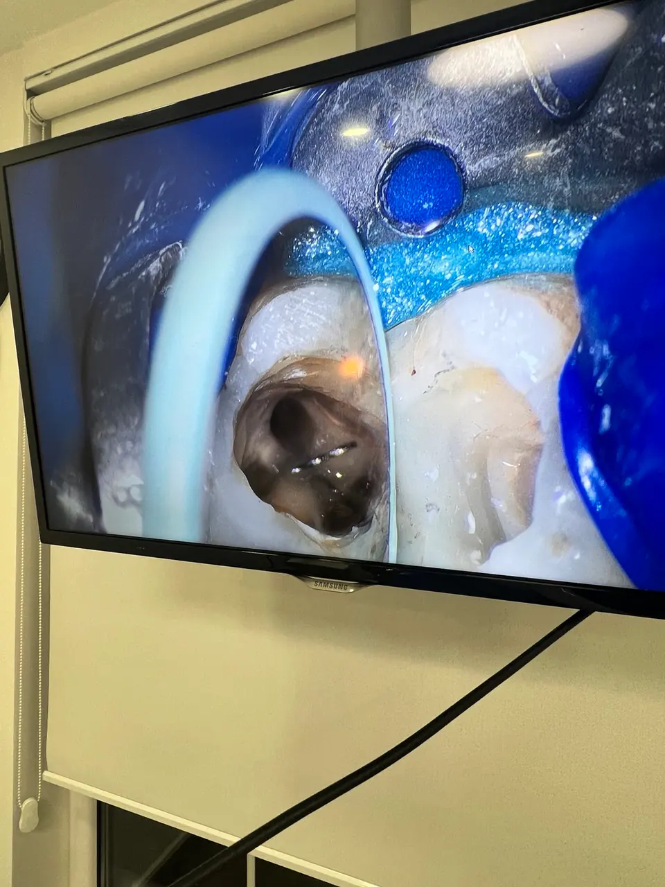
Magnified endodontic working field under an operating microscope

What does the microscope add?

The operating microscope enlarges the working field and directs light along the clinician’s viewing axis. This can support controlled access, identification of canal entries and inspection of the pulp chamber floor.

When can it be particularly useful?

Examples include complex anatomy, calcified canals, removal of previous materials, separated instruments and suspected cracks. The benefit depends on the specific tooth and procedure.

Why does it not guarantee success?

Visibility is only one component. Diagnosis, isolation, disinfection, restoration, patient factors and follow-up also affect the prognosis of a tooth.

Appointment and assessment

Your treatment plan
starts with an examination.

We listen to your concerns and expectations, complete the necessary clinical assessment and explain appropriate options clearly.

Message us on WhatsApp
Call WhatsApp