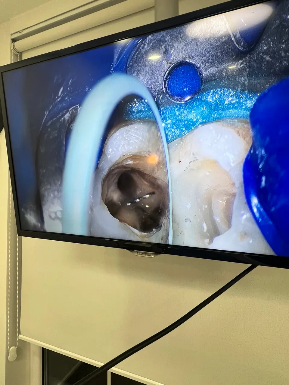
When may a microscope be used?
Magnification and illumination may assist when locating additional canals, negotiating narrow or calcified canals, removing previous filling materials, managing separated instruments or assessing a suspected crack.
Not every tooth requires the same approach. Microscope use is considered alongside examination and imaging findings.
Approach to retreatment
If symptoms persist or a lesion does not heal after previous root canal treatment, the possible cause is investigated first. The coronal restoration, quality of the root filling, possible cracks and tissues around the root are assessed together.
When retreatment is appropriate, existing materials are removed in a controlled manner, the canal system is disinfected again and the long-term restorative plan is reviewed.
A microscope is one part of treatment
Magnification improves visibility, but it does not guarantee an outcome by itself. Accurate diagnosis, isolation, disinfection, material selection, restoration and follow-up remain essential parts of care.