Initial assessment
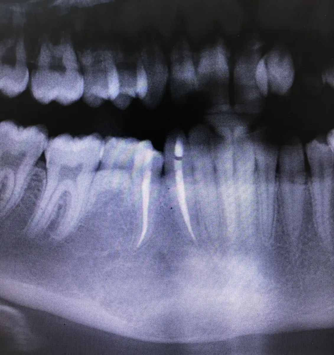
The radiograph showed an extensive radiolucent area extending through the mandibular anterior region and symphysis, involving the root areas of teeth 31–46. The patient had been referred for root canal treatment of the teeth associated with the lesion before the planned surgical excision.
Diagnosis is more than the size of the image
Each tooth within the lesion area was assessed clinically and with vitality tests. Teeth 43 and 44 were found to be non-vital, so endodontic treatment was limited to these two teeth. Teeth that responded to vitality testing were not root-treated.
A large lesion does not mean that every adjacent tooth requires root canal treatment.
A conservative endodontic approach
The initial objective was to control the source of infection, monitor the lesion and reduce the potential extent of surgical intervention. Clinical findings and radiographic changes were assessed together during follow-up.
Approximately 12 months of follow-up
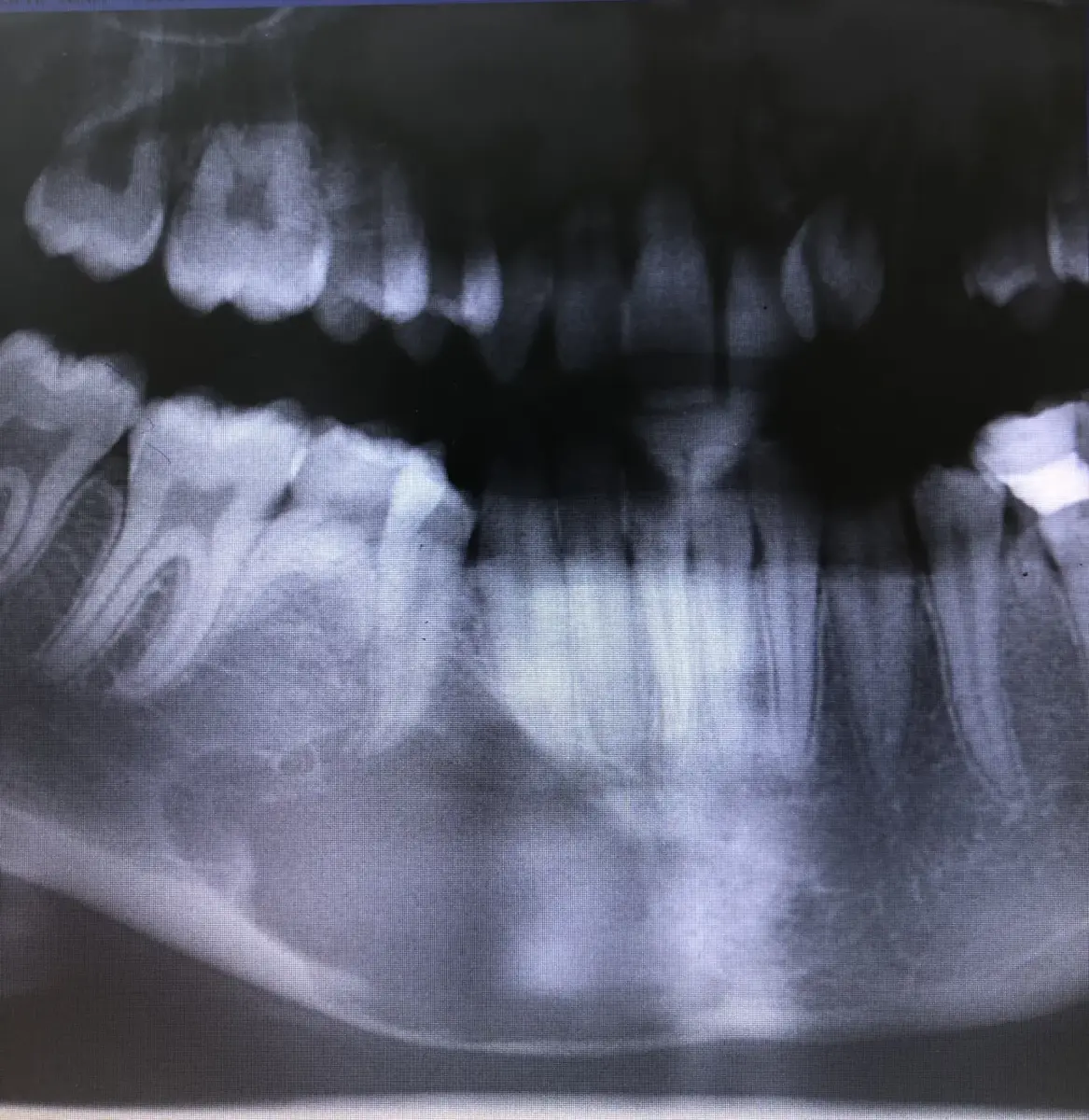
There were 345 days between the baseline and review images. Complete radiographic healing was observed on the follow-up radiograph; in this individual case, the patient no longer required surgery.
This outcome relates to a single clinical case. It does not mean that every lesion with a similar appearance will heal in the same way; diagnosis and treatment decisions require an individual clinical assessment.